
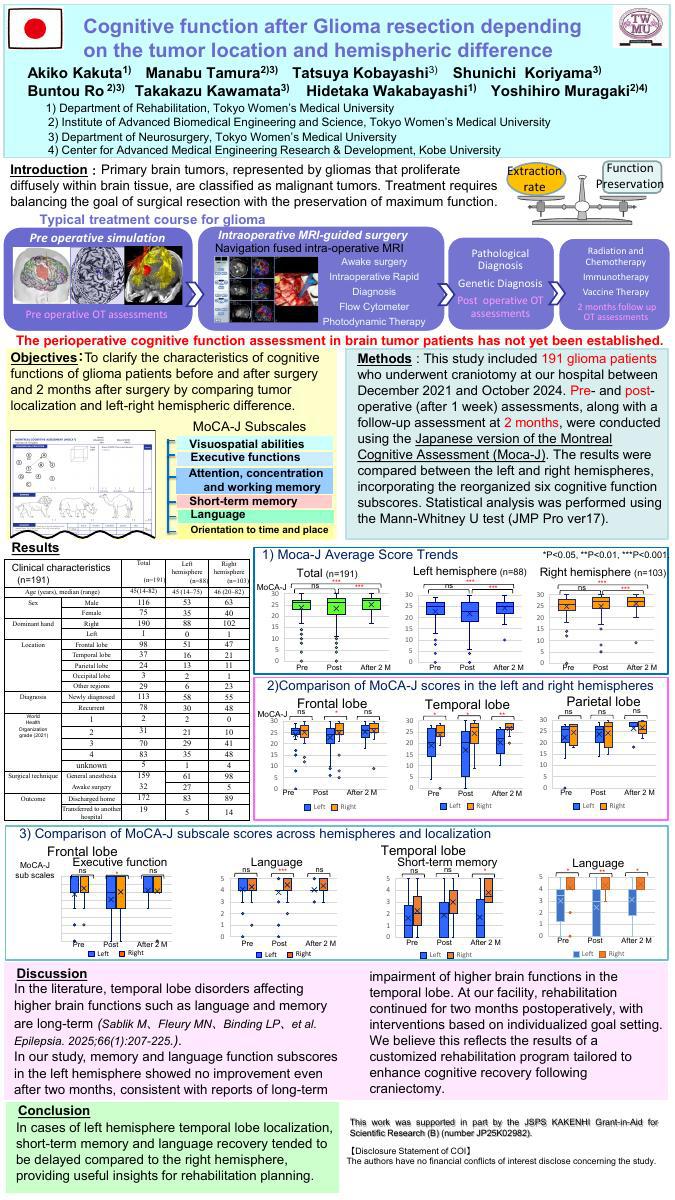
Introduction: Perioperative cognitive function in glioma patients have not yet been evaluated.
Objective: To clarify the characteristics of cognitive functions of glioma patients before and after surgery and 2 months after surgery by comparing tumor localization and left-right hemispheric difference.
Methods: We studied 198 glioma patients who underwent craniectomy at our hospital from December 2021 to October 2024. The patients were assessed with the Japanese version of the Montreal Cognitive Assessment (Moca-J) before and after surgery and 2 months later, and were compared with 6 reorganized cognitive function sub-scores in terms of localization in the left and right hemispheres. Tests were statistically analyzed with the Mann-Whitney U test (JMP pro ver17).
Results: The Moca-J mean score decreased from 22.7 to 21.7 in the left hemisphere after surgery, but recovered to 24.6 after 2 months, while the right hemisphere remained unchanged (24.7, 25.0, 26.3). Sub scores at 2 months showed that short-term memory and language were lower in the left temporal lobe than in the right temporal lobe. The sub-scores after 2 months were lower in the left temporal lobe cases than in the right temporal lobe cases.
Conclusion: The left hemisphere tumors, especially those localized in the temporal lobe, tend to have lower and more prolonged cognitive function in short-term memory and language compared to the right, which may provide useful suggestions for planning rehabilitation.