
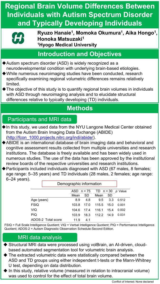
Introduction: Autism spectrum disorder (ASD) is widely recognized as a neurodevelopmental condition with underlying brain-based etiologies. While numerous neuroimaging studies have been conducted, research specifically examining regional volumetric differences remains relatively limited.
Objectives: The objective of this study is to quantify regional brain volumes in individuals with ASD through neuroimaging analysis and to elucidate structural differences relative to TD individuals.
Methods: We analyzed MRI data from the NYU Langone Medical Center, obtained via the Autism Brain Imaging Data Exchange (ABIDE). ABIDE is an international, publicly accessible neuroimaging repository aggregating MRI data from multiple universities and research institutions. The acquisition and use of these data were approved by the institutional review boards of the respective contributing institutions. Participants included individuals diagnosed with ASD (67 males, 8 females; age range: 5–35 years) and TD individuals (28 males, 2 females; age range: 6–24 years). Structural MRI data were processed using volBrain, an AI-driven, cloud-based automated segmentation tool for volumetric brain analysis. The extracted volumetric data were statistically compared between the ASD and TD groups using either independent t-tests or the Mann-Whitney U test, depending on data distribution.
Results: The ASD group showed a significantly smaller right orbital part of the inferior frontal gyrus and larger right inferior temporal and fusiform gyri than the TD group.
Conclusion: The orbital part of the inferior frontal gyrus is critically implicated in social cognition, while the inferior temporal gyrus and fusiform gyrus are key regions associated with language processing and facial recognition. The observed volumetric differences in these regions suggest potential neuroanatomical correlates underlying social and communication impairments in ASD.